
Figure 1 Article Yushun Wan
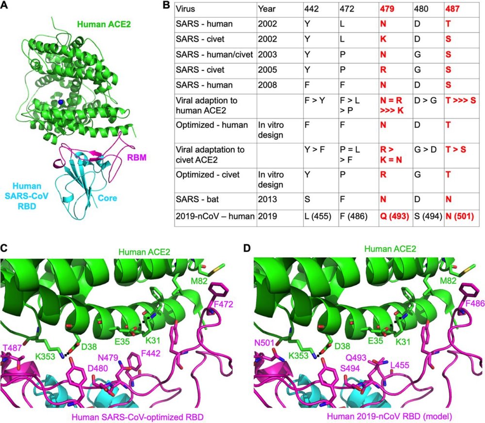
Fig1 - Structural analysis of human ACE2 recognition by 2019-nCoV and SARS-CoV.
(A) Overall structure of human SARS-CoV RBD (year 2002) complexed with human ACE2. PDB ID is 2AJF. ACE2 is in green, the core of RBD (receptor-binding domain) is in cyan, and RBM (receptor-binding motif) is in magenta.
(B) Critical residue changes in the RBMs of SARS-CoV and 2019-nCoV. All these five residues in SARS-CoV underwent natural selections and were shown to be critical for ACE2 recognition, cell entry, and host range of SARS-CoV. The residue numbers are shown as in SARS-CoV RBD, with the corresponding residue numbers in 2019-nCoV shown in parentheses. Information about the two most critical residues, 479 and 487, is in red.
(C) Experimentally determined structure of the interface between a designed SARS-CoV RBD (optimized for human ACE2 recognition) and human ACE2. PDB ID is 3SCI.
(D) Modeled structure of the interface between 2019-nCoV RBD and human ACE2. Here, mutations were introduced to the RBD region in panel C based on sequence differences between SARS-CoV and 2019-nCoV.
GenBank accession numbers are MN908947.1 for 2019-nCoV spike, NC_004718.3 for human SARS-CoV spike (year 2002; strain Tor2), AGZ48818.1 for bat SARS-CoV spike (year 2013; strain Rs3367), AY304486.1 for civet SARS-CoV spike (year 2002; SZ3), and AY525636 for human/civet SARS-CoV spike (year 2003; strain GD03). References for the other sequences are in parentheses as follows: civet SARS-CoV spike (year 2005) (9); human SARS-CoV spike (year 2008) (8).